A study conducted on the skeletal differences of the knees among people, showed that there were differing average sized dimensions between the sexes and between different ethnic groups; generally the men of all ethnicities within the study were approximately 5mm larger on the front and back of the knee than the female measurements (Mahfouz et al 172). On average African American women had a patella groove that was deeper by 7.4mm, among other differences compared to caucasian females (Mahfouz et al 172). For me, this study shows that it is normal for there to be a range of size differences and skeletal differences among human beings; some knees will be stronger, some weaker and some smaller or larger. This highlights the importance of understanding that with adjustments and alignments in the yoga classroom, it cannot be a one-size-fits-all approach. Encouraging the students to listen and be present within their bodies, with the assistance of techniques such as focusing upon the breath, may be a great way to facilitate the prevention of injuries.
According to Goni (4346), a significant postural distortion is knock knees, where the knees make contact with each other when standing in a neutral position, while the distance between the ankles continues to expand; this creates problems with both running and walking; the root cause normally being overweight, Rickets, one weaker leg or some kind of chronic illness. Some of the recommended asanas to help correct this problem include Vriksasana, Padmasana, Baddha Konasana and Upavista Konasana (Goni 4347-8). This is because these postures help to rotate the knee externally and help to bring the leg back into alignment - with regular practice. Knock knees (or varus) should not be confused with averagely aligned knees; generally the medial part of the knee is bigger and wider than the lateral part which may make the knees appear as though they are not facing forward in poses such as Downward-facing-dog; the cue here should never be to face the knees to the front, as this may cause injury (Richards online).
A protective layer of articular cartilage encapsulates the extremities of the tibia and femur, as well as the underneath of the patella (Stephens 66). Another layer of fibrocartilage intra-articular pads (the medial and lateral meniscus) provide additional protection by acting as a suspension system, which prevents direct impact against one another (Stephens 66). Medial meniscus tears happen frequently in the yoga classroom; damage may be caused entirely within the class or could be the result of stress upon an old injury (Stephens 66). One such posture that may cause problems is Padmasana where there is risk of imposing rotation at the hip before the hip is ready which in turn transmits strain into the knee joint; as these joints don't receive much life-giving blood, they heal at a slow pace (Stephens 66). I think it is important to recognise here that the body is all connected, as one whole entity, as is demonstrated in this example where tension in the hips translates to knee problems. I also find it necessary to recognise the importance of gently breathing into the postures, and working our way slowly towards the full posture, over time, opposed to forcing our way in too soon.
A number of ligaments aid in bringing a stabilizing action to the knee; they are stretched when the leg is straight (Stephens 66). A standing asana that provides great stability is Tadasana because it provides maximum connection between the tibia and femur; if the knees are locked or hyperextended, however, injuries may occur; plenty people do this without even knowing it, and what happens is the anterior menisci is unduly compressed, forcing the flesh into an unnatural position; for many, the knees should be reconditioned into a moderate and relaxed alignment (Richards online). This is a range of motion difference that you may find among students. While hyperextended knees is not considered natural or healthy motion, some individuals can and do hyperextend their knees far beyond what is considered normal due to previous injuries or stress on the ligaments. In order to prevent further injury the yoga teacher should look for this, and correct the posture quickly and gently by asking the student to rest standing in slight flexion, working on quadricep strength to help the control of the knee and prevent worsening of the tendon and ligament laxity (Sean Colio online).
With the knee bent, the ligaments are softened, which permits some rotation in the knee in asanas like Padmasana (Stephens 66). Flexion in the knee in poses such as Virabhadrasana II, also creates less stability, because of a smaller surface area of contact between the two connecting bones of the leg (Richards online). With less stability in the bony structure, the ligaments and supporting muscles are doing more work and are at greater risk (Richards online). On the sides of the knee you find the medial and lateral collateral ligaments (MCL and LCL), which restrict sideward movement, and are aided in this task by muscles that extend over them (Stephens 67). The MCL preserves the integrity of the medial edge of the knee, while the LCL safeguards the lateral part of the knee (Stephens 67). Within the joint of the knee are two ligaments: the anterior cruciate ligament and the posterior cruciate ligament (ACL and PCL) which form a cross (Stephens 67). The ACL joins the tibia to the femur in the middle of the knee; it inhibits gyration of the knee and prevents the femur from sliding off the front of the tibia (Stephens 67). The PCL can be found behind the ACL and prevents extravagant rearwards movement of the knee articulation (Stephens 67). In lunging postures such as Virabadrasana I and II, the ACL is fundamental in creating stability, and is also at risk if the body is misaligned (Stephens 67). Proper alignment and the principle of steadiness and ease is essential in preventing injury here.
According to a study, the knee’s normal range of motion should allow for flexion of about 133 to 153 degrees and should be able to extend entirely straight; anything less than this is considered to be a “limited range of motion’’ (O’Connell online). Some diseases can limit the range of motion of our joints, including arthritis and cerebral palsy, among others; ageing can also affect range of motion (O’Connell online). A study showed that the practice of yoga was able to increase knee extension by 28%, and muscular endurance in knee flexion increased 57%, during an 8 week yoga program (Tran et al 166-7). This suggests that there is much hope for those who are suffering with range of motion limitations due to disease and age. Some of the postures used included: Vajrasana and Ardha Padmasana (which personally I am cautious of with fussy knees); Parsva Uttanasana, which I feel is excellent for everyone as long as the knees are not locked; three rounds of Surya Namaskara; Vrksasana and Trikonasana among others (Tran et al 166-7).
The very strong patella ligament is often referred to as the patellar tendon, since there isn’t a distinct segregation in the connection of the quadriceps tendon - which encompasses the patella, and the region where the patella and tibia join together (Stephens 67). This ligament provides the knee with power and efficiency and acts as a cover for the femur’s condyles (Stephens 67). The muscles involved with the functioning of the knee include: the adductors, the abductors, the quadriceps, the sartorius and the hamstrings - these muscles assist the ligaments of the knee in stabilisation (Stephens 67). The lateral stability of the knee is assisted by the abductor muscles, the gluteus and tensor fascia latae join to the iliotibial band, which connects to the lateral tibial condyle under the knee (Stephens 67). The functionality of the gracilis (adductor), sartorius (flexion and lateral rotation) and one of the hamstrings, the semitendinosus provide a stabilising action upon the medial part of the knee; they attach from the medial part of the tibia below the knee and run up to various other parts of the pelvis or iliac spine (Stephens 67-68). Both the lateral and medial stabilisers of the knee also assist in the rotation of the tibia, when the knee is in flexion in a pose such as Vrksasana (Stephens 68-69).
The strongest muscles participating in knee extension, flexion and stabilisation, are the hamstrings and quadriceps; the quadriceps, being the strongest, has four heads all joining to one part, creating the quadriceps tendon, which expands over the forepart of the knee to convert into the patellar tendon and attaches onto the patella’s proximal side; through the connection to the patellar tendon the quadriceps action is transmitted to the tibia (Stephens 69). Of the four heads of the quadriceps, three come from the femoral shaft: vastis medialis, vastus lateralis, vastus intermedius; the fourth, the rectus femoris, originating from the upper frontal part of the pelvis, plays an important part in the extension of the knee and flexion of the hip as can be sampled in Utthita Hasta Padangusthasana (Stephens 69). The combined strength of these muscles are supported by the formation of the patella which acts like a pivot point (Stephens 69). The concentric contraction of the quadriceps (which causes the muscles to shorten) or their isometric contraction (which causes the muscles to create resistance without lengthening or shortening), creates extension of the knee and a stretching in the hamstrings in some seated and standing asanas; the eccentric contraction (causing a lengthening of the muscles), adds to the elevation of the physique in back-bending asanas (Types of Muscle Contractions Online & Stephens 69).
According to Richards (online), the vastus medialis (located inside the front thigh), is predominantly accountable for maintaining the position of the kneecap in the femoral sulcus - a channel at the extremity of the femur - which should preferably glide effortlessly upward and downward within that channel. This smooth function makes the fulcrum-like action efficient in flexion and extension motion (Richards online). The vastus medialis is smaller and weaker than the vastus lateralis which is on the outer side of the front thigh; this disparity of power in the quadriceps, may result in the kneecap pulling up and out, resulting in affliction when walking or in lunging asanas (Richards online). Richards (online) recommends the following exercise in order to create steadiness in relation to this issue: take a rolled towel and place it beneath your knees while sitting in Dandasana; flex your feet with your toes pointing upwards while pushing your heels away from your body; push your knees downwards allowing your inner knee to lead the way for 10-30 seconds and then let go and redo this action until tired.
The knees’ primary flexors are the hamstrings; on the medial part of the knee are the semimembranosus and the semitendinosus which arise from the ischial tuberosity; they provide support for the knee medially and help with medial circular motion; on the lateral side of the knee we get support and stability from the biceps femoris (also a hamstring), which arises from the rear of the ischial tuberosity, and the rear of femoral shaft; the two origins conjoin, before moving over the lateral part of the knee, and then attaching to the proximal fibula (Stephens 70).
While at first glance the knee may seem to be a simple hinge joint, a deeper look reveals many complexities beneath the surface. The knees are an important joint that receive impact from the hips and feet, allowing us to move, walk and run; a vital factor in many yoga asanas. It would appear that when your knees cause you to beg for mercy, a conscious yoga practice can be of great usefulness upon the healing journey, by increasing strength and range of motion. On the other hand (or knee), if one is too rushed to cultivate proper alignment and connect with the breath, truly listen to your body or the bodies of your students, then the knees can be at great risk of gaining new injuries or evoking old ones.
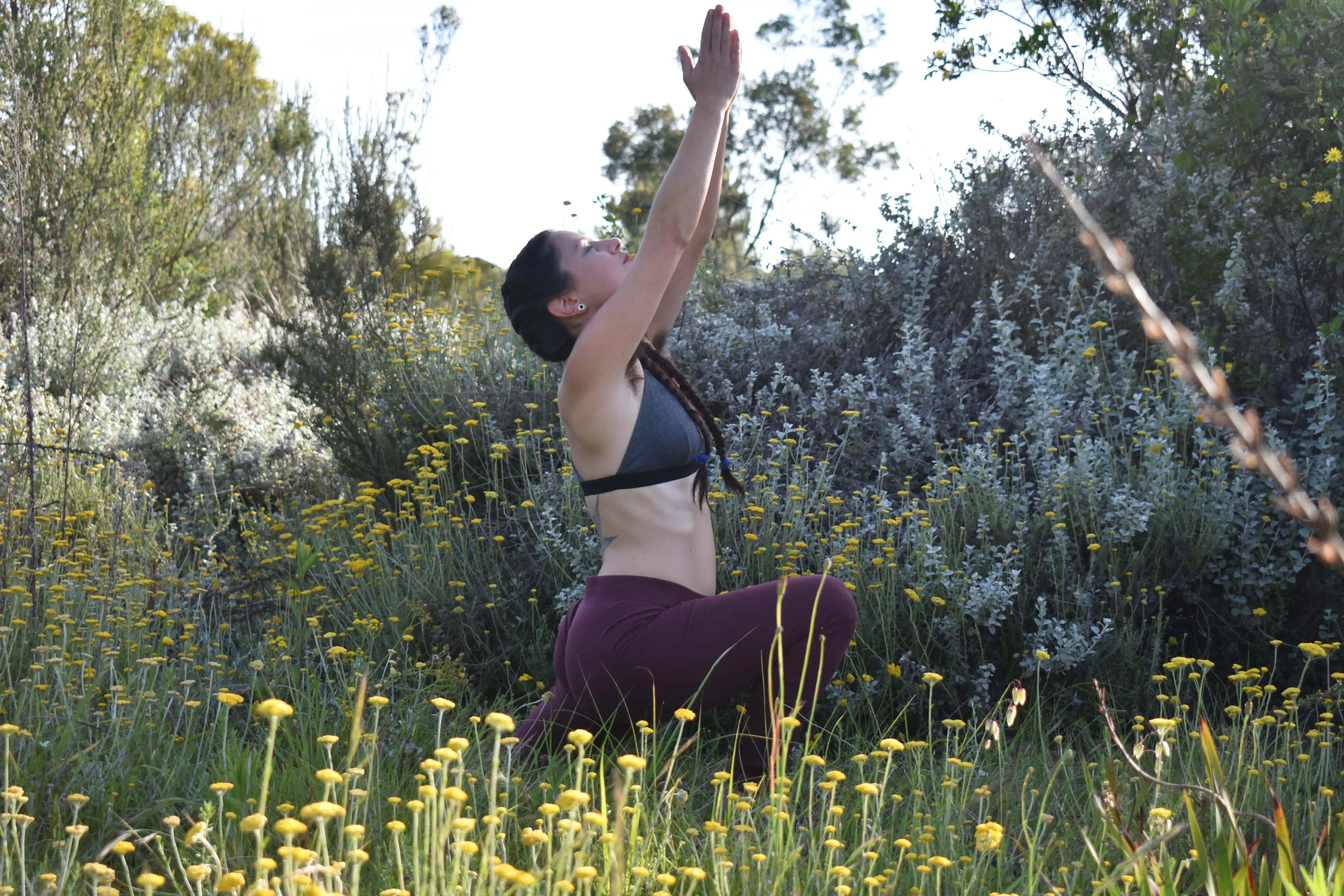
Photo captured by Gheon Steencamp
References: check these formats
Goni, O. “Treatment of Common Deformities Through Yoga”. International Journal of Social Science and Economic Research, Vol 3, no 8, 2018, Pp. 4337-4353. Ijsser. ISSN: 2455-8834.
Mahfouz, Mohamed et al. “Three-Dimensional Morphology of the Knee Reveals Ethnic Differences”. Clinical Orthopaedics & Related Research, vol 470, no. 1, 2011, pp. 172-185. Ovid Technologies (Wolters Kluwer Health), doi:10.1007/s11999-011-2089-2.
Mansfield, P. and Neumann, D. Essentials of kinesiology for the physical therapist assistant. 3rd ed. St. Louis, Mo.: Mosby. 2009. Pp. 279-298.
O’Connell, K. “What is Limited Range of Motion?”. Healthline. 2019. https://www.healthline.com/health/limited-range-of-motion.
Richards, M., 2019. How to Keep Your Knees Safe and Injury-Free During a Yoga Class. Yoga Journal, [online] Available at: <https://www.yogajournal.com/teach/anatomy-yoga-practice/keep-your-knees-safe/> [Accessed 10 May 2021].
Sean Colio, MD. “Understanding Knee Hyperextension”. Sports-Health, 2021, https://www.sports-health.com/sports-injuries/knee-injuries/understanding-knee-hyperextensi on.
Stephens, Mark. Teaching Yoga Essential Foundations and Techniques. North Atlantic Books. 2010. Pp.66-70.
Tran, MD., Holly, RG et al. “Effects of Hatrha Yoga Practice On the Health-Related Aspects of Physical Fitness”, Preventative Cardiology, vol 4, no. 4, 2007, pp. 165-170. Wiley, doi:10.1111/j.1520-037x.2001.00542.x.
Types of Muscle Contractions: Isotonic and Isometric. 14 Aug. 2020, https://med.libretexts.org/@go/page/7549.


